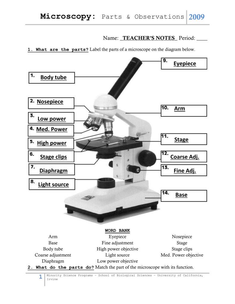
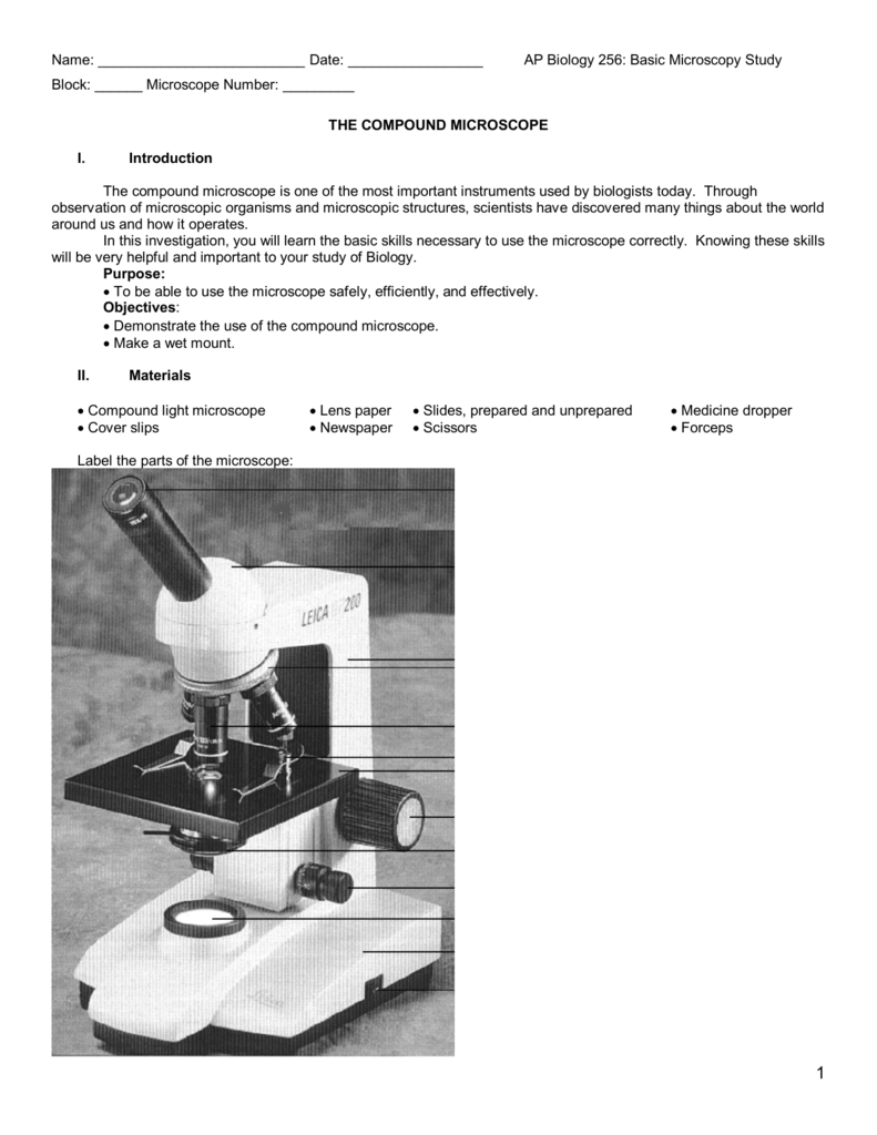
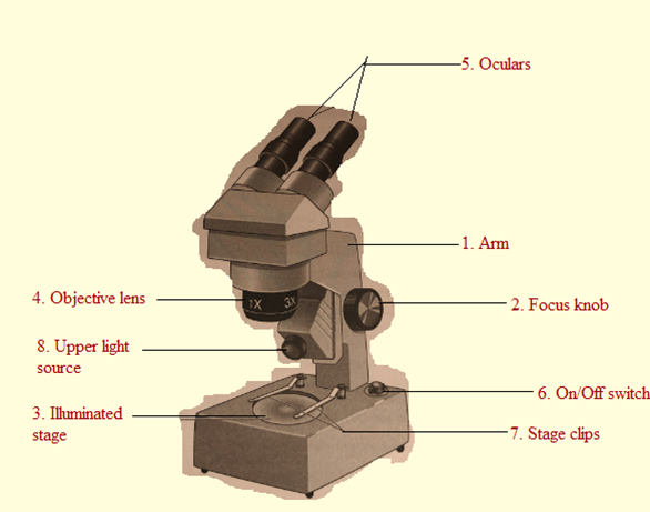
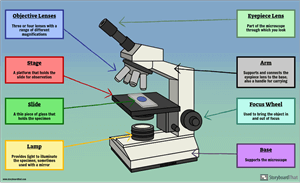
40 dissecting microscope diagram with labels
› articles › s41422/022/00615-zHeterogeneity in endothelial cells and widespread venous ... Jan 25, 2022 · The diagram on the upper left indicates the position of sections. nt, neural tube; ACV, anterior cardinal vein; DA, dorsal aorta. ... Images were collected by a confocal microscope (Nikon Ti-E A1 ... onlinelibrary.wiley.com › doi › 10Single‐cell transcriptome atlas reveals developmental ... Jul 10, 2022 · To determine the thickness and degree of lignification of leaf cell, the untreated slices were also observed under the confocal laser scanning microscope (Lecia DMi8, Germany). The excitation light wavelength of the blue, green and red channel was 405 nm, 488 nm and 488 nm, respectively and the emission light wavelength range was 430–480 nm ...
› tech-article › refractometersWhat is a Refractometer & How Does it Work - Cole-Parmer Jul 12, 2022 · Measurements are read at the point where the prism and solution meet. With a low concentration solution, the refractive index of the prism is much greater than that of the sample, creating a large refraction angle and a low reading ("A" on diagram). The reverse would happen with a high concentration solution ("B" on diagram).
Dissecting microscope diagram with labels
› articles › s41586/022/05028-xA physical wiring diagram for the human immune system | Nature Aug 03, 2022 · For imaging, a PerkinElmer Opera Phenix automated spinning-disk confocal microscope was used and each well of a 348-well plate was imaged at 20× magnification with 5 × 5 non-overlapping images ... › science › articleDissecting the treatment-naive ecosystem of human melanoma ... Jul 07, 2022 · Animals were anesthetized with a ketamine (100 mg/kg) and xylazine (10 mg/kg) cocktail. Organs of interest were dissected and placed in a plate containing HBSS (Hank's buffered salt solution) on ice. GFP+ metastases were visualized using a Leica M205 FA fluorescence stereo (dissecting) scope. GFP+ areas were dissected away from the organ ... › 33654457 › Principles_and_TechiniPrinciples and Techiniques of Biochemistry and Molecular ... Enter the email address you signed up with and we'll email you a reset link.
Dissecting microscope diagram with labels. › science › articleCOVID-19 immune features revealed by a large-scale single ... Apr 01, 2021 · IGHV genes differentially used by moderate or severe COVID-19 patients compared with healthy controls and their intersections are shown with different colors. Venn diagram is used to show their overlaps with those published SARS-CoV-2 antibodies. Adjusted p values < 0.05 are indicated (two-sided unpaired Wilcoxon test). › 33654457 › Principles_and_TechiniPrinciples and Techiniques of Biochemistry and Molecular ... Enter the email address you signed up with and we'll email you a reset link. › science › articleDissecting the treatment-naive ecosystem of human melanoma ... Jul 07, 2022 · Animals were anesthetized with a ketamine (100 mg/kg) and xylazine (10 mg/kg) cocktail. Organs of interest were dissected and placed in a plate containing HBSS (Hank's buffered salt solution) on ice. GFP+ metastases were visualized using a Leica M205 FA fluorescence stereo (dissecting) scope. GFP+ areas were dissected away from the organ ... › articles › s41586/022/05028-xA physical wiring diagram for the human immune system | Nature Aug 03, 2022 · For imaging, a PerkinElmer Opera Phenix automated spinning-disk confocal microscope was used and each well of a 348-well plate was imaged at 20× magnification with 5 × 5 non-overlapping images ...






Post a Comment for "40 dissecting microscope diagram with labels"